Incidence of early pseudo-progression in a cohort of malignant glioma patients treated with chemoirradiation with temozolomide

Incidence of Early Pseudo-progression in a Cohortof Malignant Glioma Patients Treated WithChemoirradiation With Temozolomide BACKGROUND. Radiotherapy (RT) plus concomitant and adjuvant temozolomide (TMZ) is now the standard of care for patients with newly diagnosed glioblas- toma. The occurrence of pseudo-progression directly after RT is a recognized phenomenon, but to the authors’ knowledge its incidence after combined RT/ TMZ is unknown. The occurrence of early pseudo-progression was retrospectively assessed in a cohort of malignant glioma patients treated with RT/TMZ.
METHODS. The pre-RT and post-RT brain scans from patients treated with RT/ TMZ for a malignant glioma were reviewed. Scans were made before the start of RT, 4 weeks after the end of RT, and every 3 months thereafter. In addition, infor- 1 Department of Neuro-Oncology/Neurology, Eras- mation was collected regarding clinical signs and symptoms, dexamethasone mus MC/Daniel den Hoed Cancer Center, Rotter- RESULTS. Eighty-five patients were identified. In 36 patients (42%) the first follow- 2 Department of Radiology, Erasmus MC/Daniel up scan 4 weeks after the end of RT indicated disease progression. Of these 36 den Hoed Cancer Center, Rotterdam, The Nether- patients, 18 (50%) were diagnosed with pseudo-progression. None of the patients received additional treatment other than TMZ. Six of 18 patients with pseudo-pro- 3 Department of Radiotherapy, Erasmus MC/ gression and 12 of the 18 patients with real tumor progression developed new clini- Daniel den Hoed Cancer Center, Rotterdam, The cal signs and symptoms during RT or in the first 4 weeks thereafter.
CONCLUSIONS. Up to 50% of malignant glioma patients treated with RT/TMZ and 4 Department of Radiotherapy, University Medical progression immediately after RT develop pseudo-progression. The current study Center Utrecht, Utrecht, The Netherlands.
data support the idea to continue TMZ in the case of progressive lesions immedi- ately after RT/TMZ. Surgery should be considered in symptomatic cases. The inclusion of patients with progressive lesions developing directly after chemora- diation in studies regarding recurrent gliomas will lead to an overestimation of the results. Cancer 2008;113:405–10. Ó 2008 American Cancer Society.
Dr. van den Bent has acted as a paid consultant KEYWORDS: glioblastoma, glioma, temozolomide, radiotherapy, chemoirradiation, for Schering-Plough and is also a member of the The first 2 authors contributed equally to thiswork.
Radiotherapy (RT) and concomitant temozolomide (TMZ) fol- lowed by adjuvant TMZ has become the standard of care for patients with glioblastomas since the European and Canadian ran- Dr. Brandsma’s current address: Department of domized trial was published in 2005.1 Despite the improved out- Neuro-oncology, Netherlands Cancer Institute, come with combined modality treatment, the overall outcome of this disease remains dismal, with many patients progressing earlyafter RT or during adjuvant TMZ chemotherapy. In recent years, we Address for reprints: Walter Taal, MD, Departmentof Neuro-Oncology/Neurology, Erasmus MC/Daniel and other clinicians observed the occurrence of progressive mag- den Hoed Cancer Center, Groene Hilledijk 301, netic resonance imaging (MRI) lesions immediately after the end 3075 EA Rotterdam, The Netherlands; Fax: (011) of concurrent chemoirradiation with TMZ (RT/TMZ), with spon- 31-10-4391031; E-mail: [email protected] taneous improvement without further treatment other than adju- Received December 11, 2007; revision received vant TMZ. In an earlier study, this phenomenon of early pseudo- February 18, 2008; accepted March 4, 2008.
progression was investigated in 32 patients with a malignant glioma DOI 10.1002/cncr.23562Published online 16 May 2008 in Wiley InterScience (www.interscience.wiley.com).
who received RT only.2 In that study, 3 of 9 patients (33%) with a progressive lesion immediately after RT Characteristics at Baseline of 85 Patients With Malignant demonstrated a stabilized or improved lesion during at Glioma Treated With Radiotherapy Plus Concomitantand Adjuvant Temozolomide least 6 months on subsequent scans, without addi-tional treatment. To our knowledge, the incidence of early pseudo-progression in malignant glioma patients treated with RT/TMZ is unknown, nor is it known whether this is clinically symptomatic. (At the time of publication, Brandes et al have since published data regarding the incidence of pseudo-regression and the correlation with the MGMT promoter methylation sta- tus in glioblastoma patients.3) We investigated the inci- Complete or partial resection vs biopsy, % dence of early pseudo-progression and its clinical WHO indicates World Health Organization.
features by reviewing a cohort of patients with newlydiagnosed malignant gliomas who were treated withRT plus concomitant and adjuvant TMZ.
Early disease progression was defined as progres- sion (25% increase) noted on the MRI scan 4 weeksafter RT and concomitant TMZ, with or without neu- rologic deterioration, and on a stable or higher dose For this study, all malignant glioma patients treated of dexamethasone. Real early progression was scored between 2000 and July 2006 with RT/TMZ in the if the patient with early progression developed addi- Daniel den Hoed Cancer Center in Rotterdam, The Netherlands, were reviewed. Furthermore, all patients 6 months. Pseudo-early progression was scored if the who participated in the European Organization for patient with early progression 1) had at least a 50% Research and Treatment of Cancer (EORTC) 22981 decrease in the enhancing lesion during further fol- study and received chemoirradiation at the Univer- low-up, while remaining neurologically stable and on sity Medical Center Utrecht in Utrecht, The Nether- a stable or decreasing dose of dexamethasone (a ‘partial response’ according to the criteria of Macdo- excluded. Treatment was comprised of fractionated nald et al.4) or 2) remained clinically and radiologi- irradiation at a dose of 2.0 grays (Gy) per fraction cally stable with a stable or decreased dosage of given once daily on weekdays over a period of steroids for at least 6 months after RT/TMZ without 6 weeks to a total dose of 60 Gy and concomitant any further treatment other than adjuvant cycles of TMZ (75 mg/m2/day on all days), followed after 4 TMZ. Clinical features of the patients with real early weeks by 6, 28-day cycles of adjuvant TMZ (Day 1–5 progression and pseudo-early progression were com- every 28 days, at a dose of 150–200 mg/m2/day).
pared. Kaplan–Meier survival curves were used to an- None of the patients received other treatment, such as gliadel or a focal radiotherapy boost.
progression and pseudo-early progression.
Clinical records were reviewed concerning the type of surgery performed, histology, radiation field, Eighty-five patients were treated with RT plus concomi- dose, and survival. Per treatment protocol, brain ima- tant and adjuvant TMZ. The majority of the patients had ging was performed before RT (median interval of a glioblastoma multiforme (GBM). Table 1 summarizes 14 days between the brain scan and the initiation of the demographic and clinical features of these patients.
RT), 4 weeks after the end of RT (median interval of In 39 patients, the pre-RT/TMZ scan was a computed 30 days between the end of the RT/concomitant tomography (CT) scan; in the other 46 patients, MRI TMZ), and thereafter every 3 months. Two independ- was used. All follow-up scans were MRI scans.
ent reviewers reviewed all brain scans (HGdB and Thirty-six of the 85 patients (42%; 95% confi- WT). The evaluation was based on precontrast and dence interval [95% CI], 31.5–52.5%) were identified postcontrast images and primarily on the changes in as having early progression on the first follow-up the contrast-enhancing area. In the case of disagree- scan 4 weeks after RT and concomitant TMZ com- ment, the scans were jointly re-evaluated. The pared with the pre-RT imaging (Table 2). Thirty-one response criteria developed by Macdonald et al.4 were of the 68 patients (45%; 95% CI, 33.2–56.8%) with a used to quantify all changes in the enhancing lesions GBM and 5 of the 17 patients (29%; 95% CI, 7.4– on the scan, clinical status, and dexamethasone dose.
50.6%) with an anaplastic glioma had early disease Pseudo-progression in Glioma Patients/Taal et al.
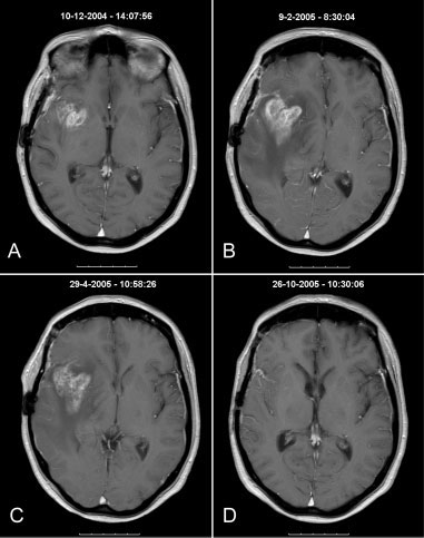
progression. In only 1 patient could the decreaseddose of dexamethasone explain the observed pro-gression noted on the MRI scan (Patient 17). Threepatients did not continue with adjuvant TMZbecause of neurologic deterioration (Patients 22, 31,and 34). All of the remaining patients continued withadjuvant TMZ.
Eighteen of 36 patients (50%; 95% CI, 33.7– 66.3%) with early disease progression were diagnosedwith pseudo-early progression. Pseudo–early progres-sion was noted in 15 of the 31 patients (48%; 95% CI,30.4–65.6%) with a GBM and in 3 of the 5 patients(60%; 95% CI, 17.0–100%) with an anaplastic glioma.
In 17 of the 36 patients with early disease progres-sion, the enhancing lesion on subsequent MRI scansstabilized for at least 6 months (Patients 4, 5, 11, 13,14, and 15), decreased (Patients 2, 6, 7, 8, 9, 12, 16,and 18) (Fig. 1), or disappeared completely (Patients1, 10, and 17). Because these 17 patients were alsoclinically stable or improved and were receiving astable or decreasing dose of dexamethasone, theywere scored as having pseudo-early progression. Oneof the 36 patients with early progression underwenta re-resection 3 months after RT and concomitantTMZ because of further deterioration; at surgery,only necrosis was found (Patient 3). The patient con- FIGURE 1. Patient 8 was a 50-year-old female who was diagnosed with a tinued with 3 more cycles of adjuvant TMZ and right temporal glioblastoma. Debulking surgery was followed by radiotherapy remained stable for another 15 months.
and concomitant temozolomide (RT/TMZ). Compared with (A) the pre-RT/TMZ To investigate whether the percentage of patients scan, the (B) scan of the brain taken 4 weeks after RT/TMZ demonstrated with pseudo-early progression was artificially high disease progression within the area of gadolinium uptake. The patient because of the use of a CT scan rather than an MRI remained clinically stable and was receiving a stable dose of dexametha- scan before RT, we separately analyzed the patients sone. She continued with 3 cycles of adjuvant TMZ and the dexamethasone diagnosed with early disease progression who had dose was gradually lowered and withdrawn. (C) The magnetic resonance been evaluated with MRI scans both before and after imaging (MRI) scan taken 3 months after RT/TMZ was unchanged and she RT. In this group, 8 of 17 patients (47%) were subse- received another 3 cycles of TMZ. (D) The MRI scans taken 6 months and 9 quently diagnosed with pseudo-early progression months, respectively, after RT/TMZ demonstrated a diminishing lesion. The compared with 10 of 19 patients (53%) who had a patient developed disease progression 12 months after RT/TMZ.
The individual charts of the patients with pseudo-early progression were re-examined for other pseudo–early progression (Table 2; P 5 .313, chi- explanations of disease remission, but none were square) The volume of the radiation field was not found. In particular, no new treatments had been found to be significantly different between the patients with real early progression, pseudo-early Neurologic deterioration was found in 6 of the progression, and no early progression (data not 18 patients (33%) with pseudo-early progression and shown). Pseudo–early progression was observed in 6 in 12 of the 18 patients (67%) with real early progres- of 26 patients who underwent a biopsy (23%; 95% sion during RT or in the first 4 weeks thereafter. The CI, 6.8–39.2%) and in 13 of 59 patients who under- mean age of the patients with real early progression went a partial or complete surgical resection (22%; was significantly higher compared with that of the 95% CI, 11.4–32.6%) The extent of surgical resection patients with pseudo–early progression (55 years vs could not be taken into consideration because no 46 years, respectively; P 5 .0342). The World Health direct postoperative scans were made.
Organization (WHO) performance status was not The survival curves of the patients with early dis- found to be significantly different between the ease progression (split between those with pseudo– patients with real early progression and those with early progression and those with real early progres- Pseudo-progression in Glioma Patients/Taal et al.
concomitant TMZ. Fifteen of these 26 patientsunderwent surgery again and 7 of them were diag-nosed with radiation necrosis.
Although we hypothesized that pseudo–early progression would occur more frequently after RT/concomitant TMZ compared with RT only, the inci-dence we observed (18 of 85 patients [21%]; 95% CI,12.5–29.9%) is still within the range de Wit et al.2observed after the use of RT only (3 of 32 patients[9%]; 95% CI, 20.7–19.5%). One possible explanationfor the increased awareness of the phenomenon ofpseudo–early progression could be that most GBMpatients are now treated with RT/TMZ and thereforeare more closely followed. Conversely, in cell lines,synergy between TMZ and RT has been demon-strated in MGMT promoter gene methylated tumors.7 FIGURE 2. Kaplan-Meier survival curves of malignant glioma patients trea- It may well be that this synergistic antitumoral effect ted with chemoirradiation with temozolomide. Survival curves of patients causes more profound tumor necrosis and inflamma- developing disease progression 4 weeks after radiotherapy and concomitant tion with vascular changes, leading to a deficient temozolomide, split into patients with further disease progression (real early blood-brain barrier mimicking enhancing tumor on a progression), patients who remained stable for at least 6 months or scan. Further research will explore whether there is improved (pseudo early progression), and patients without early disease pro- indeed a correlation between MGMT methylation sta- gression (no early progression), are shown.
tus and the occurrence of pseudo–early progression.
(At the time of publication, Brandes et al have since sion) and patients with no early progression are published data regarding the incidence of pseudo- regression and the correlation with the MGMT pro-moter methylation status in glioblastoma patients.3)The increase in radiation necrosis noted to occur if chemotherapy is given after RT in patients with brain In the past decades, the sporadic occurrence of early tumors also suggests that more intensified treat- ments cause more severe local reactions.7 This is abnormalities immediately after RT with spontaneous also the likely explanation for the earlier occurrence recovery have been described.5 Since the introduc- of radiation necrosis noted after combined chemoir- tion of chemoirradiation with TMZ for GBM, there radiation with TMZ.6,8 Most likely, pseudo–early pro- has been an increasing awareness of this phenom- enon. From our cohort of 85 patients treated with continuum, with more severe local reactions leading chemoirradiation with TMZ, the progressive en- to new focal signs and symptoms and true radiation hancement was not found to be because of tumor progression in 18 of the 36 patients (50%) with a pro- The precise mechanism of this early post-RT/ gressive lesion at the time of first tumor evaluation concomitant TMZ deterioration is unknown. The after chemoirradiation. Although the pre-RT/con- underlying mechanism may be varied and in addi- comitant TMZ scan was a CT scan in approximately tion to the above-mentioned radiation-induced (and half of these patients, it is unlikely that this influ- perhaps vascular endothelial growth factor [VEGF] enced the results because the outcome was the same signaling-dependent) vascular and necrotic changes, in patients who underwent an initial MRI scan. In tumor progression during the first part of RT and addition, the patients with pseudo-early progression subsequent response could also be an explanation.
were found to have a similar survival compared with Although to our knowledge the exact nature of patients without early progression (Fig. 2). A recent this pseudo-early progression is unknown, these report examining surgery performed within 6 months observations have important consequences for trials from RT/concomitant TMZ in patients with GBM of recurrent malignant glioma. Of the 36 patients in corroborated our findings regarding the frequency of the current study with early progression (according nontumoral increase in enhancement.6 In that study, to the response criteria of Macdonald et al.), 3 26 of 51 GBM patients demonstrated disease progres- achieved a complete response and 8 achieved a par- sion within 6 months after the completion of RT/ tial response, whereas 6 fulfilled the criteria for stable disease at 6 months. Six-month progression-free sur- then, we advise continuing adjuvant TMZ in patients vival (PFS) is currently considered the most valid with early disease progression and not to include endpoint for phase 2 studies of recurrent GBM.9 If these patients in studies of malignant gliomas that the presently reported patients with immediate dis- recur within 3 months after RT/TMZ. Surgery should ease progression would all have been entered in a be considered in the case of patients who develop phase 2 trial of recurrent GBM, this would have lead early clinical signs and symptoms and a progressive to a false-positive study result with a 6-month PFS lesion. If mainly or only necrosis is found at the time of surgery, treatment with TMZ should be continued.
From the clinical perspective, an important question is how to differentiate between pseudo–early progression and real early progression immedi-ately after RT. Because of the inherent risks and inva- siveness of a stereotactic biopsy, it is not very Stupp R, Mason WP, van den Bent MJ, et al. Radiotherapyplus concomitant and adjuvant temozolomide for glio- attractive to obtain histologic proof of these lesions.
blastoma. N Engl J Med. 2005;352:987–996.
Moreover, it is not clear whether all cases with de Wit MC, de Bruin HG, Eijkenboom W, Sillevis Smitt pseudo-early progression will show only necrosis at PA, van den Bent MJ. Immediate post-radiotherapy biopsy because tumor cells may still be present. Clin- changes in malignant glioma can mimic tumor progres- ical deterioration during or within the 4 weeks after Brandes AA, Franceschi E, Tosoni A, et al. MGMT pro- RT/concomitant TMZ cannot be used to distinguish moter methylation status can predict the incidence and between these entities because clinical deterioration outcome of pseudoprogression after concomitant radio- was also observed in the group of patients with chemotherapy in newly diagnosed glioblastoma patients.
pseudo-early progression, although less frequently (33% vs 67%). WHO performance score, biopsy ver- Macdonald DR, Cascino TL, Schold SC Jr, Cairncross JG.
Response criteria for phase II studies of supratentorial sus surgical resection, and volume of the radiation malignant glioma. J Clin Oncol. 1990;8:1277–1280.
field also cannot be used to discriminate between Griebel M, Friedman HS, Halperin EC, et al. Reversible pseudo–early progression and real early progression.
neurotoxicity following hyperfractionated radiation ther- The median age in the group of patients with real apy of brain stem glioma. Med Pediatr Oncol. 1991;19: early progression was found to be higher (56 years vs Chamberlain MC, Glantz MJ, Chalmers L, Van Horn A, 46 years), which could simply reflect the higher like- Sloan AE. Early necrosis following concurrent Temodar lihood of disease progression in elderly patients.
and radiotherapy in patients with glioblastoma. J Neu- Again, this finding is also of no value in individual patients. Furthermore, pseudo–early progression was Chakravarti A, Erkkinen MG, Nestler U, et al. Temozolo- also noted in 3 of 17 patients with anaplastic glioma mide-mediated radiation enhancement in glioblastoma: areport on underlying mechanisms. Clin Cancer Res.
(17%; 95% CI, 20.9–34.9%) versus 15 of 68 GBM patients (22%; 95% CI, 12.2–31.9%).
Ruben JD, Dally M, Bailey M, Smith R, McLean CA, Fedele To our knowledge, to date it has been unclear P. Cerebral radiation necrosis: incidence, outcomes, and whether modern imaging techniques such as posi- risk factors with emphasis on radiation parameters and chemotherapy. Int J Radiat Oncol Biol Phys. 2006;65:499–508.
spectroscopy, or diffusion-weighted and perfusion Ballman KV, Buckner JC, Brown PD, et al. The relationship imaging can be used to make a distinction, although between 6-month progression-free survival and 12-month some data appear to be promising.10 Patients with overall survival end points for phase II trials in patients real early progression have a terrible outcome asso- with glioblastoma multiforme. Neuro Oncol. 2007;9:29–38.
ciated with continuing standard TMZ (Fig. 2); future Zeng QS, Li CF, Liu H, Zhen JH, Feng DC. Distinctionbetween recurrent glioma and radiation injury using mag- efforts to better identify these patients are critical so netic resonance spectroscopy in combination with diffu- that a potential opportunity to salvage them with an sion-weighted imaging. Int J Radiat Oncol Biol Phys.
alternative therapeutic intervention is not lost. Until

Source: http://www.tnrd.org.tr/files/downloads/makaleler/w_taal_pseudoprogresyon_incidence.pdf

Microsoft word - dermedicaconsultcard.docx

Name _________________________________________________________________________________________________ Address _______________________________________________________________________________________________ _________________________________________________________________________________________________ Phone ______________________________________________________ email: ________________________

emr.moh.gov.rw

Code review /ProgramOverModule11Purpose of the module: the module displays statistics about a program. All available programs can be selected. The list of indicators is static. The user can select to display a graph. Maintainability - JUnit AdminListExtensionTest fails, no other test cases implemented - Design: org.openmrs.module.programOver.web.controller.ProgramPatientsController is not a Spr

Copyright © 2009-2018 Drugs Today